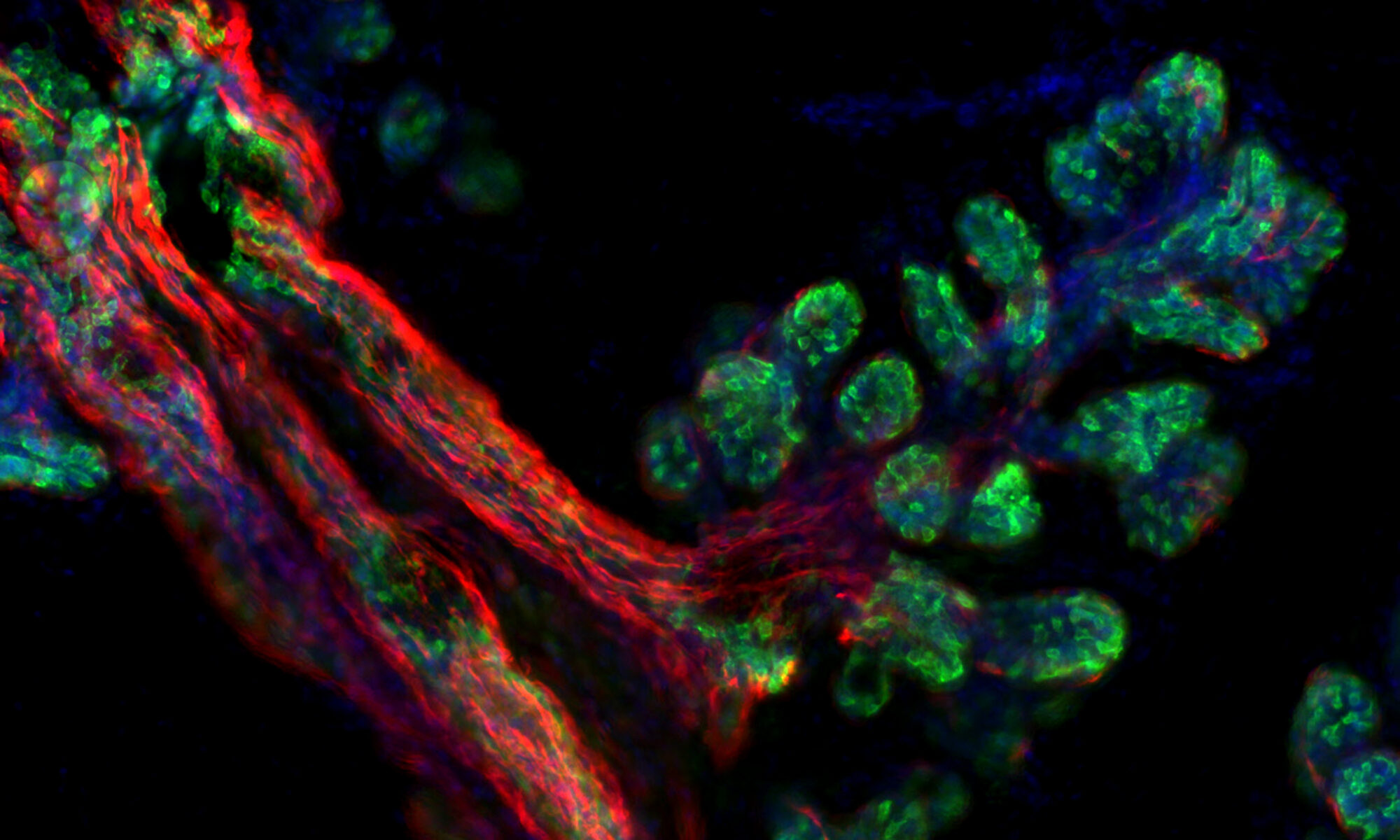
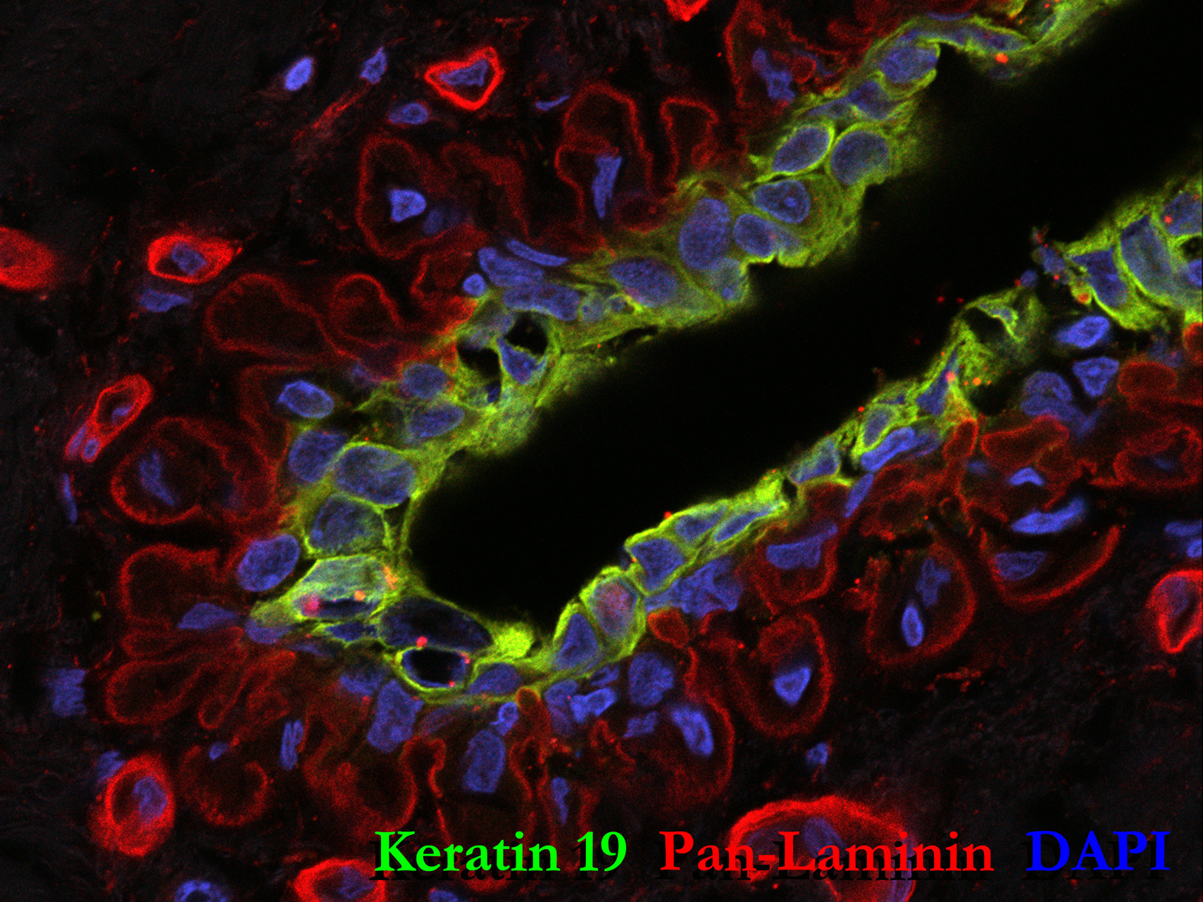
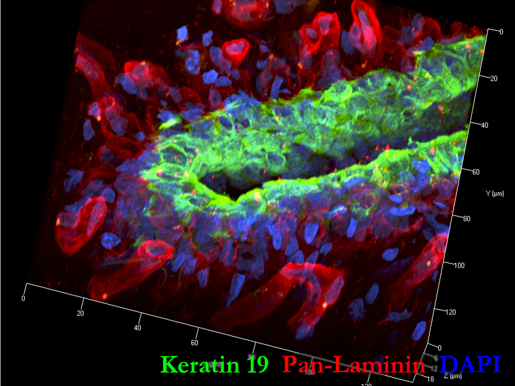
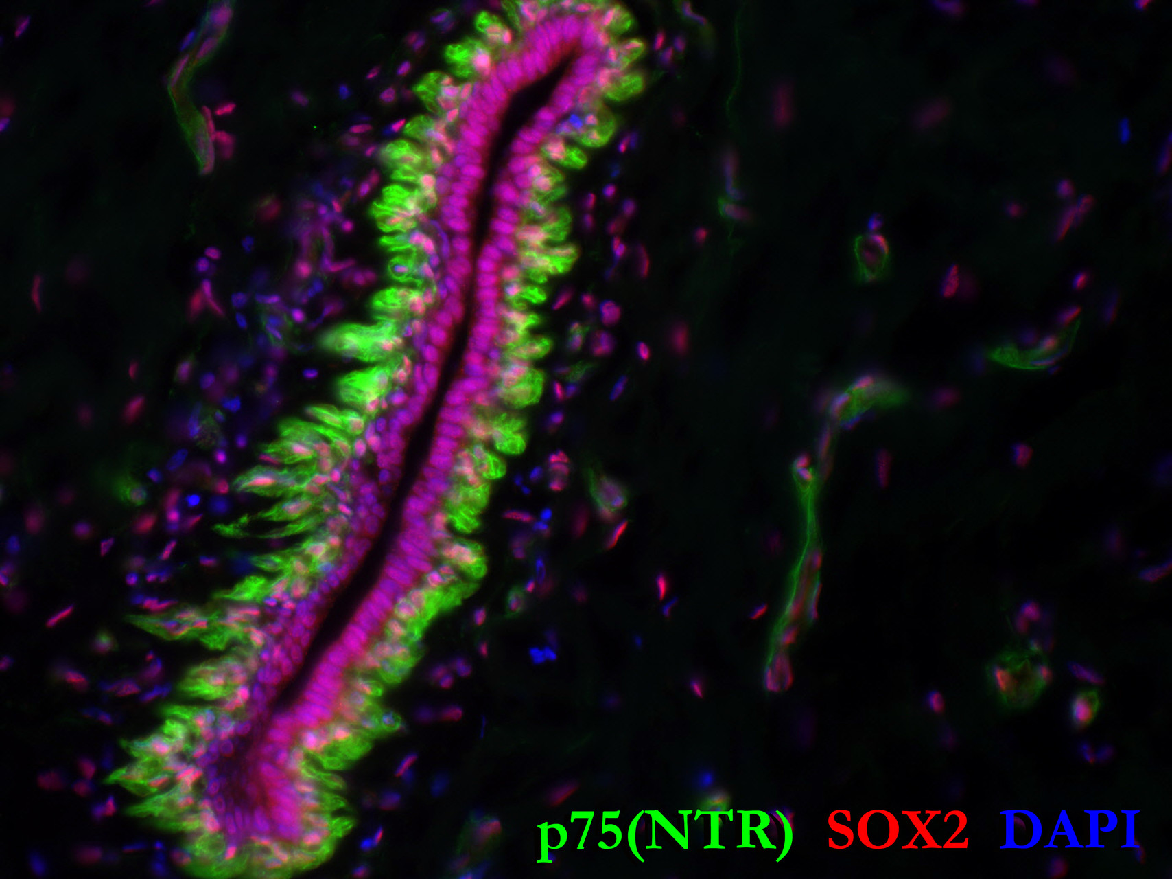

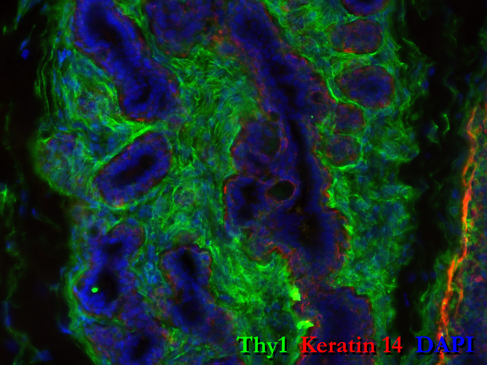
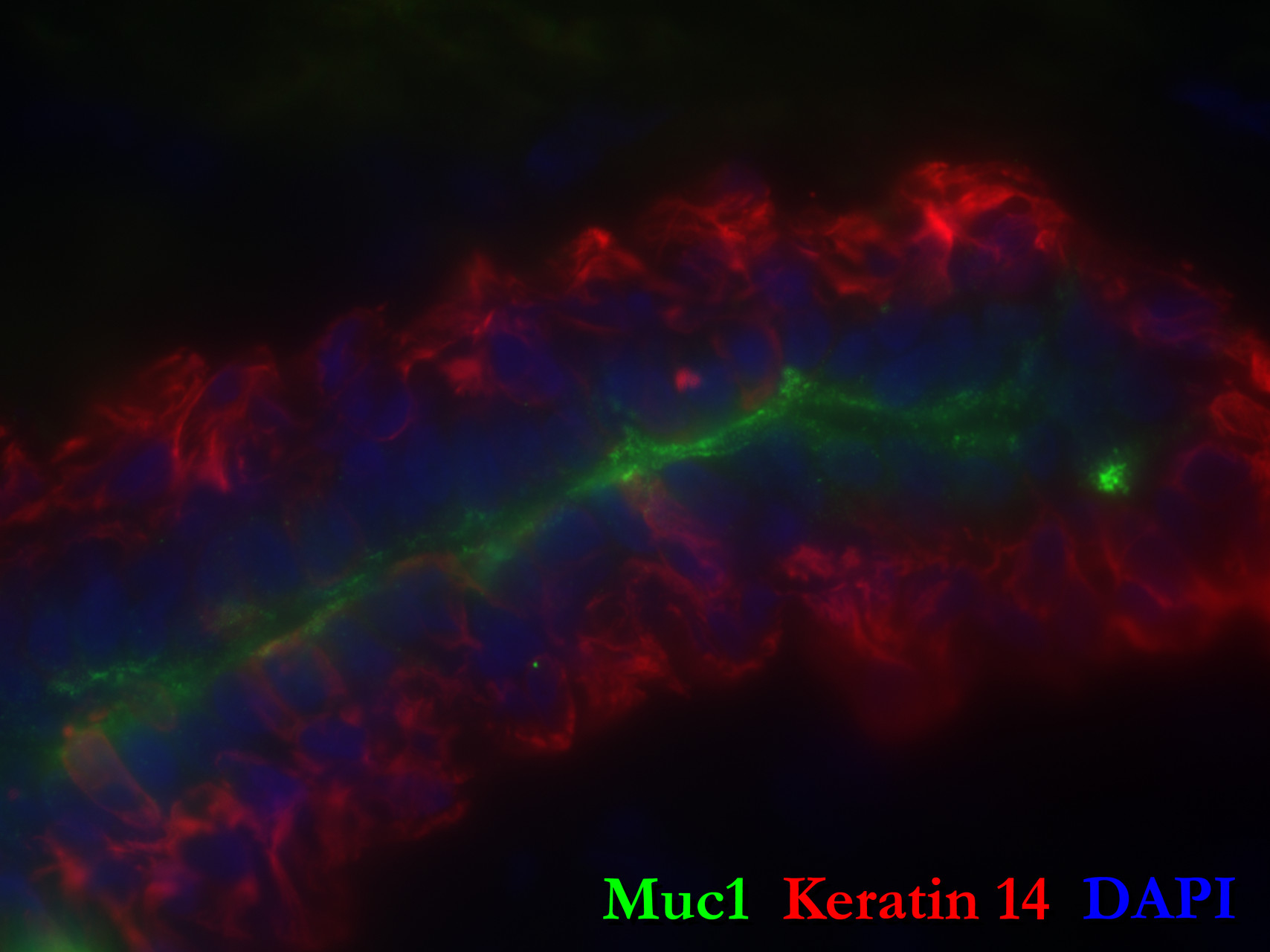
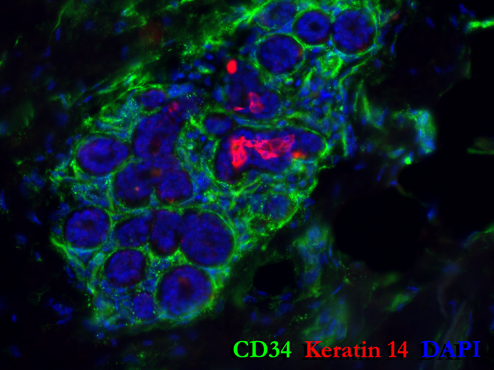
The above images are immunostained tissues of normal breast, using a variety of antibody combinations to reveal aspects of the tissue. These images highlight the wonderfully complex architecture and beauty of the composition of the tissue.
A larger collection of images can be found on Google Photos:
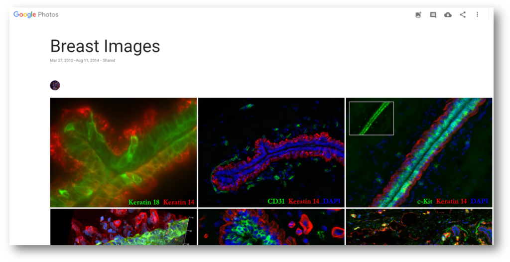
Stained with over 50+ antibodies, our collection consists of hundreds and hundreds of images. . . and counting -as we add to it daily. We’ve stained these tissues to decipher the types and spacial orientation of the cell types composing the breast; and to also guide development of a flow cytometry method to sort and isolate the different cell types for experimental study. The bulk of images are not publicly available yet, but we hope to share them soon (after acceptance of our manuscript) So in the meantime, please enjoy the dozen or so we have posted.
Here, Curt describes immunostaining to U.S. Representative George Miller during his 2012 visit to Lawrence Berkeley National Lab:
